Macular Degeneration
Age-related Macular Degeneration (AMD) is the leading cause of blindness and loss of central vision among adults over the age of 65. AMD is a chronic disease that affects the retina – the part of the eye that allows you to see fine detail — blurring your central vision. It can have devastating impact on your ability to read, drive, or engage in other activities of daily life. An estimated 15 million North Americans alone have AMD.
There are two forms of AMD, known as “dry” and “wet”:
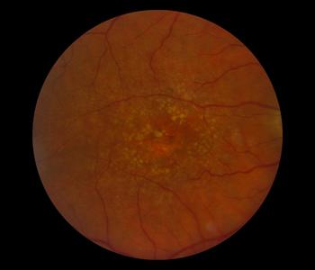
- Dry AMD – characterized by small yellow particles and pigment changes in the macula caused by cells breaking down. Over time, the deposits may grow together and harden, thereby interfering with central vision.
- Wet AMD – Occurs when abnormal blood vessels behind the retina start to grow under the macula. These blood vessels often leak blood and fluid. Wet AMD is the more advanced form of the disease and loss of vision occurs quickly.
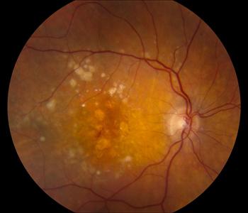
Early Stage Wet AMD
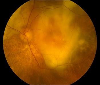
End Stage Wet AMD
Most patients have what is called the “dry” form, in which the yellow deposits, called drusen, are present in the macula – in some patients eventually leading to a dimming or distortion of vision. In advanced stages, tissue death may lead to blind spots and loss of central vision. About 10 percent of people with AMD develop the “wet” form of the disease in which abnormal blood vessels grow.
Symptoms
The earliest signs of macular degeneration in the retina can be detected before you have any vision loss. This detection is facilitated by an eye exam in which eye drops are given in a physician’s office, which dilate the pupil of the eye.
Patients with macular degeneration are usually older than 55 years old, have signs of macular degeneration in both of their eyes and may have experienced some slow, insidious vision loss. Most have dry macular degeneration. Perhaps 85 – 90% of macular degeneration is of the “dry” variety.
Dry AMD usually occurs slowly, over time. The patient may notice a need for brighter light when reading. Other symptoms may include difficulty adapting to low light levels, increased blurriness of printed words, decrease in brightness of colors, or a blurred spot in the center of the field of vision. A dark blank or black spot in the middle of your vision can also be a sign of macular degeneration. This spot starts out small, grows over time and could eventually lead to legal blindness in your central vision but does not affect your peripheral or side vision. So you never become totally blind.
In contrast, visual changes in wet AMD occur more rapidly, resulting in an abrupt decline in central vision. Patients may experience visual distortions, such as straight lines appearing wavy, or objects appearing larger or smaller than they are. As in dry AMD, patients may also notice a well-defined blind spot in the center of vision.
You should also keep an eye out for early symptoms of macular degeneration. Here are some things to look for:
Watch for straight lines that appear broken, crooked, wavy, bent or distorted in your vision. One way to test your eye sight is to use an Amsler Grid.
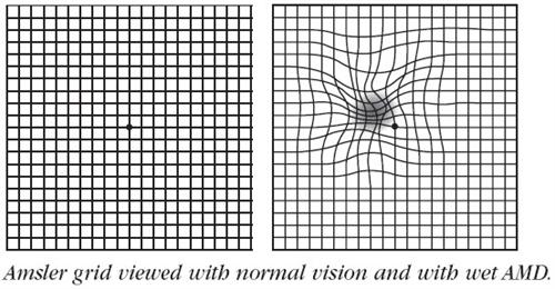
If the lines in the grid appear anything but straight and unbroken, you might be experiencing a typical symptom of macular degeneration.
However, this test is hardly sufficient to rule out the possibility that one has developed macular degeneration. Many people with macular degeneration may see no abnormalities on an Amsler grid, so don’t use this as a substitute for regular good dilated eye examinations of the retina.
Pay close attention to a decline in your central vision, both close-up and distant. People use central vision when they drive, read, look at faces or view pictures. Your central vision allows you to see details, colors and shapes more clearly.

Visual acuity is often reduced and the patchy vision that results often interferes with visual efficiency.
Regular dilated eye exams with an ophthalmologist are important, especially when you’re at higher risk for macular degeneration. If you are over the age of 50, an exam every one to two years is a good idea in order to look for signs of macular degeneration before any vision loss has occurred.
Causes
AMD is caused by the destruction of light-sensitive cells in the macula, the central portion of the retina in the back of the eye. The light-sensitive cells of the macula give us our ability to have sharp, detailed vision.
In a healthy eye, images are focused onto the retina and then converted into electrical signals that are sent to the brain for processing.
During normal aging, yellowish deposits, called drusen, form under the retina. It is possible to have drusen with no accompanying loss of vision.
But as drusen increase in size and number, they can interfere with proper functioning of the retina, damaging or killing the light-sensitive cells of the macula. This is how dry AMD occurs.
The wet form of AMD occurs when blood vessels behind the retina begin to grow in an abnormal way. These newly formed blood vessels can then leak blood and fluid, causing the macula to swell. Again, the macula’s light-sensitive cells are damaged or killed.
Risk factors
The primary risk factor for AMD is age. The older you are, the greater your risk for macular degeneration. Also, people with a family history of the disease are at higher risk for macular degeneration, as are women, and people of European descent.
Complications
Dry AMD is the more common and less severe form of AMD, but is often a precursor to wet AMD. Although approximately 80 percent of patients with AMD have the dry form, the wet form is responsible for 80 to 90 percent of severe loss of vision or legal blindness associated with AMD.