Fluorescein Angiography
Angiography is a diagnostic procedure in which a rapid sequence of photographs are taken to document the blood circulation of the retina. In this test, illuminated dye is injected into the body through your veins (IV), usually in the arm, forearm or hand. As your blood flows, the dye gradually appears in the retina.
Since the fluorescein dye is a very bright yellow, the skin may appear jaundiced for a few hours and then the yellow color disappears. The dye is excreted through the kidneys causing the urine to be a bright yellow for 24-36 hours. The coloration of this dye is considered to be a normal result of the after effects.
I will photograph the retina and evaluate its appearance with the help of the illuminated dye. This analysis helps determine if the disease is present and how far it has progressed.
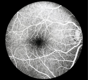
Normal eye as seen in fluorescein angiography
 |
 |
as seen in fluorescein angiography
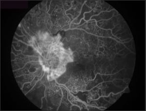
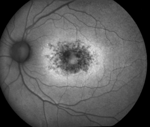
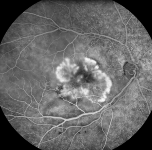
Diabetic Retinopathy under Fluorescein Angiography